The presence of poor esophageal body function can impact the likelihood of relief of regurgitation, dysphagia, and respiratory symptoms following surgery and may influence the decision to undertake a partial rather than a complete fundoplication. When peristalsis is absent or severely disordered, many would opt for a partial fundoplication, although recent studies would suggest a complete fundoplication may be appropriate even in this setting. The less favorable response of atypical, compared with typical, reflux symptoms after fundoplication may be related to persistent poor esophageal propulsive function and the continued regurgitation of esophageal contents.
The function of the esophageal body is assessed with esophageal manometry. This is performed with five pressure transducers located in the esophagus. To standardize the procedure the most proximal pressure transducer is located 1 cm below the well-defined cricopharyngeal sphincter. With this method a pressure response along the entire esophagus can be obtained during one swallow. The study consists of recording ten standard wet swallows with 5 mL of water. Amplitude, duration, and morphology of contractions following each swallow are all calculated at the five discrete levels within the esophageal body. The delay between onset or peak of esophageal contractions at the various levels of the esophagus is used to calculate the speed of wave propagation and represents the degree of peristaltic activity.
Esophageal disorders are frequently associated with abnormalities of duodenogastric function. Symptoms suggestive of gastroduodenal pathology include nausea, epigastric pain, anorexia, and early satiety. Abnormalities of gastric motility or increased gastric acid secretion can be responsible for increased esophageal exposure to gastric juice. If not identified before surgery, unrecognized gastric motility abnormalities are occasionally unmasked by an antireflux procedure, resulting in disabling postoperative symptoms. Considerable experience and judgment are necessary to identify the patient with occult gastroduodenal dysfunction. The surgeon should maintain a keen awareness of this possibility and investigate the stomach given any suggestion of problems. Tests of duodenogastric function that are helpful when investigating the patient with GE reflux include gastric emptying studies, gastric acid analysis, 24-hour gastric pH monitoring, and ambulatory bilirubin monitoring of the esophagus and stomach.
Poor gastric emptying or transit can provide for reflux of gastric contents into the distal esophagus. Standard gastric emptying studies are performed with radionuclide-labeled meals. They are often poorly standardized and difficult to interpret. Emptying of solids and liquids can be assessed simultaneously when both phases are marked with different tracers. After ingestion of a labeled standard meal, gamma camera images of the stomach are obtained at 5- to 15-minute intervals for 1.5 to 2 hours.
After correction for decay, the counts in the gastric area are plotted as percentage of total counts at the start of the imaging. The resulting emptying curve can be compared with data obtained in normal volunteers. In general, normal subjects will empty 59% of a meal within 90 minutes.
Friday, March 7, 2008
 Assessment of esophageal body and gastric function

Radiographic evaluation
Radiographic assessment of the anatomy and function of the esophagus and stomach is one of the most important parts of the preoperative evaluation. Critical issues are assessed, including the presence of esophageal shortening, the size and reducibility of a hiatal hernia, and the propulsive function of the esophagus for both liquids and solids.
The definition of radiographic GE reflux varies depending on whether reflux is spontaneous or induced by various maneuvers. In only about 40% of patients with classic symptoms of GERD is spontaneous reflux observed by the radiologist (i.e., reflux of barium from the stomach into the esophagus with the patient in the upright position). In most patients who show spontaneous reflux on radiography, the diagnosis of increased esophageal acid exposure is confirmed by 24-hour esophageal pH monitoring. Therefore, the radiographic demonstration of spontaneous regurgitation of barium into the esophagus in the upright position is a reliable indicator that reflux is present. Failure to see this does not indicate the absence of disease.
A carefully performed video esophagram can provide an enormous amount of information on the structure and function of the esophagus and stomach. The modern barium swallow emphasizes motion-recording (video), utilizes a tightly controlled examination protocol, and requires an understanding of esophageal physiology.
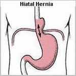
Videotaping the study greatly aids the evaluation, providing the surgeon with a real-time assessment of swallowing function, bolus transport, and the size and reducibility of hiatal hernias. Given routine review before antireflux surgery, its value becomes increasingly clear. The study provides structural information including the presence of obstructing lesions and anatomic abnormalities of the foregut. A hiatal hernia is present in more than 80% of patients with GE reflux and is best demonstrated with the patient in the prone position, which causes increased abdominal pressure and promotes distention of the hernia above the diaphragm. The presence of a hiatal hernia is an important component of the underlying pathophysiology of GE reflux. Other relevant findings include a large (greater than 5 cm) or irreducible hernia, suggesting the presence of a shortened esophagus; a tight crural collar that inhibits barium transit into the stomach, suggesting a possible cause of dysphagia; and the presence of a paraesophageal hernia.
Lower esophageal narrowing resulting from a ring, stricture, or obstructing lesion is optimally viewed with full distention of the esophagogastric region. A full-column technique with distention of the esophageal wall can be used to discern extrinsic compression of the esophagus. Mucosal relief or double-contrast films should be obtained to enhance the detection of small esophageal neoplasms, mild esophagitis, and esophageal varices. The pharynx and upper esophageal sphincter are evaluated in the upright position, and an assessment of the relative timing and coordination of pharyngeal transit is possible.
The assessment of peristalsis on video esophagram often adds to, or complements, the information obtained by esophageal motility studies. This is in part because the video barium study can be done both upright and supine and with liquid and solid bolus material, which is not true of a stationary motility examination. This is particularly true with subtle motility abnormalities. During normal swallowing, a stripping wave (primary peristalsis) is generated that completely clears the bolus. Residual material can stimulate a secondary peristaltic wave, but usually a second pharyngeal swallow is required. Motility disorders with disorganized or simultaneous esophageal contractions have tertiary waves and provide a segmented appearance to the barium column, often referred to as beading or corkscrewing. In dysphagic patients, a barium-impregnated marshmallow, bread, or hamburger is a useful adjunct, which can discern a functional esophageal transport disturbance not evident on the liquid barium study. Reflux is not easily seen on video esophagram, and motility disorders that cause retrograde barium transport may be mistaken for reflux.
Assessment of the stomach and duodenum during the barium study is a necessity for proper preoperative evaluation of the patient with GERD. Evidence of gastric or duodenal ulcer, neoplasm, or poor gastroduodenal transit has obvious importance in the proper preoperative evaluation.
Assessment of esophageal length
Esophageal shortening is a consequence of scarring and fibrosis associated with repetitive esophageal injury. Anatomic shortening of the esophagus can compromise the ability to perform an adequate tension-free fundoplication and may result in an increased incidence of breakdown or thoracic displacement of the repair. Esophageal length is best assessed preoperatively using video roentgenographic contrast studies and endoscopic findings. Endoscopically, hernia size is measured as the difference between the diaphragmatic crura, identified by having the patient sniff, and the GE junction, identified as the loss of gastric rugal folds. We consider the possibility of a short esophagus in patients with strictures or those with large hiatal hernias (greater than 5 cm), particularly when the latter fail to reduce in the upright position on a video barium esophagram.
The definitive determination of esophageal shortening is made intraoperatively when, after thorough mobilization of the esophagus, the GE junction cannot be reduced below the diaphragmatic hiatus without undue tension on the esophageal body. Surgeons performing fundoplication have reported varying incidences of esophageal shortening, attesting to the judgment inherent in defining and recognizing undue tension. An advantage of transthoracic fundoplication is the ability to mobilize the esophagus extensively from the diaphragmatic hiatus to the aortic arch. With the GE junction marked with a suture, esophageal shortening is defined by an inability to position the repair beneath the diaphragm without tension. In this situation, a Collis gastroplasty coupled with either a partial or complete fundoplication may be performed.
Potential pitfalls of laparoscopic fundoplication include the elevation of the diaphragm due to pneumoperitoneum, potentially contributing to a false impression that esophageal length is adequate, and the limited ability to mobilize the esophagus relative to the transthoracic approach. In our experience, the failure to appreciate esophageal shortening is a major cause of fundoplication failure and is often the explanation for the slipped Nissen fundoplication. In many such instances, the initial repair is incorrectly constructed around the proximal tubularized stomach rather than the terminal esophagus. Surgeons opting to perform fundoplication laparoscopically in the setting of potential esophageal shortening must be vigilant of esophageal tension, technically facile at extensive mediastinal mobilization of the esophagus while preserving vagal integrity, and able to perform a laparoscopic or open transabdominal Collis gastroplasty should esophageal lengthening be necessary.
Twenty-four hour ambulatory pH monitoring
The most direct method of assessing the relationship between symptoms and GERD is to measure the esophageal exposure to gastric juice with an indwelling pH electrode. Miller first reported prolonged esophageal pH monitoring in 1964, although it was not until 1973 that its clinical applicability and advantages were demonstrated by Johnson and DeMeester. Ambulatory pH testing is considered by many to be the gold standard for the diagnosis of GERD, because it has the highest sensitivity and specificity of all tests currently available. Some experts have suggested that 24-hour pH monitoring be used selectively, limited to patients with atypical symptoms or no endoscopic evidence of GE reflux. Given present-day referral patterns, more than half of the patients referred for antireflux surgery will have no endoscopic evidence of mucosal injury. For these patients, 24-hour pH monitoring provides the only objective measure of the presence of pathologic esophageal acid exposure. Although it is true that most patients with typical symptoms and erosive esophagitis have a positive 24-hour pH result, the study provides other useful information. It quantifies the actual time that the esophageal mucosa is exposed to gastric juice, measures the ability of the esophagus to clear refluxed acid and correlates esophageal acid exposure with the patient's symptoms. It is the only way to quantitatively express the overall degree and pattern of esophageal acid exposure, both of which may impact the decision toward surgery. Patients with nocturnal or bipositional reflux have a higher prevalence of complications and failure of long-term medical control. For these reasons, we continue to advocate its routine use in clinical practice.
The units used to express esophageal exposure to gastric juice are (a) cumulative time the esophageal pH is below a chosen threshold, expressed as the percent of the total, upright, and supine monitored time; (b) frequency of reflux episodes below a chosen threshold, expressed as number of episodes per 24 hours; and (c) duration of the episodes, expressed as the number of episodes greater than 5 minutes per 24 hours and the time in minutes of the longest episode recorded. The upper limits of normal were established at the 95th percentile. Most centers use pH 4 as the threshold. Combining the result of the six components into one expression that reflects the overall esophageal acid exposure below a pH threshold, a pH score was calculated by using the standard deviation of the mean of each of the six components measured.

