
The relief of symptoms remains the primary force driving antireflux surgery in patients with Barrett's esophagus. Healing of esophageal mucosal injury and the prevention of disease progression are important secondary goals. In this regard, patients with Barrett's esophagus are no different than the broader population of patients with GE reflux. Antireflux surgery should be considered when patient factors suggest severe disease or predict the need for long-term medical management, both of which are almost always true in patients with Barrett's esophagus.
PPI therapy, both to relieve symptoms and to control any coexistent esophagitis or stricture, is an acceptable treatment option in patients with Barrett's esophagus. Once initiated, however, most patients with Barrett's will require lifelong treatment. Complete control of reflux with PPI therapy can be difficult, however, as has been highlighted by studies of acid breakthrough while on therapy. Ablation trials have shown that mean doses of 56 mg of omeprazole are necessary to normalize 24-hour esophageal pH studies. Antireflux surgery likely results in more reproducible and reliable elimination of reflux of both acid and duodenal content, although long-term outcome studies suggest that as many as 25% of patients postfundoplication have persistent pathologic esophageal acid exposure confirmed by 24-hour pH studies.58
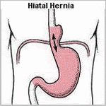
An important consideration is that patients with Barrett's esophagus generally have severe GERD, with its attendant sequelae such as large hiatal hernia, stricture, shortened esophagus, and poor motility. These anatomic and physiologic features make successful antireflux surgery a particular challenge in this population. Indeed, recent data suggest that antireflux surgery in patients with Barrett's esophagus may not be as successful in the long term as in those without Barrett's.
Studies focusing on the symptomatic outcome following antireflux surgery in patients with Barrett's esophagus document excellent to good results in 72% to 95% of patients at 5 years following surgery The outcome of laparoscopic Nissen fundoplication in patients with Barrett's esophagus has been assessed at 1 to 3 years after surgery.Reflux symptoms were absent postoperatively in 79% of the patients. Postoperative 24-hour pH was normal in 17 of 21 (81%) patients. Ninety-nine percent of the patients considered themselves cured or improved, and 97% were satisfied with the surgery.
Friday, May 23, 2008
 Treatment of Barrett's esophagus
Tuesday, May 20, 2008
What is Barrett's esophagus?
Barrett's esophagus is the metaplastic complication of Acid Reflux Disease.
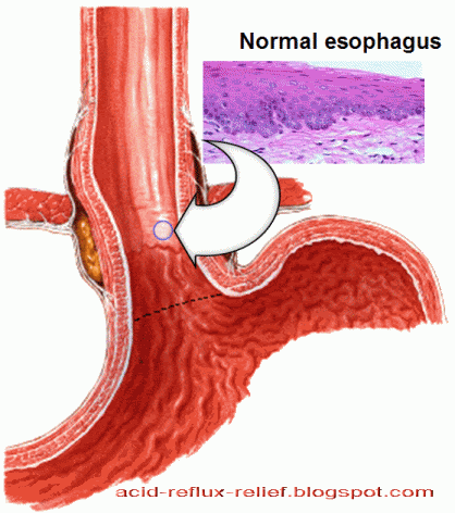
It is the condition whereby the tubular esophagus is lined with columnar epithelium rather than squamous epithelium was first described by Norman Barrett in 1950. He incorrectly believed it to be congenital in origin. It is now realized that it is an acquired abnormality, occurring in 7% to 10% of patients with GERD, and represents the end stage of the natural history of this disease. It is also understood to be distinctly different from the congenital condition in which islands of mature gastric columnar epithelium are found in the upper half of the esophagus.
The definition of Barrett's esophagus has evolved considerably over the past decade. Traditionally, Barrett's esophagus was identified by the presence of any columnar mucosa extending at least 3 cm into the esophagus. Recent data indicating that specialized intestinal-type epithelium is the only
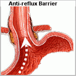
Recent studies suggest that the metaplastic process at the GE junction may begin by conversion of distal esophageal squamous mucosa to cardiac-type epithelium, heretofore presumed to be a normal finding. This is likely due to exposure of the distal esophagus to excess acid and gastric contents via prolapse of esophageal squamous mucosa into the gastric environment. This results in inflammatory changes at the GE junction or a metaplastic process, both of which may result in the loss of muscle function and a mechanically defective sphincter allowing free reflux with progressively higher degrees of mucosal injury. Intestinal metaplasia within the sphincter may result, as in Barrett's metaplasia of the esophageal body. This mechanism is supported by the finding that as the severity of GERD progresses, the length of columnar lining above the anatomic GE junction is increased.
Tuesday, August 7, 2007
How serious is Barrett's esophagus?
What do you mean by Barrett's esophagus?
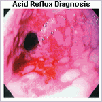
Norman Barrett (1903–1979), a British surgeon at St Thomas' Hospital, first described specified changes in the appearance of the esophageal lining of patients with acid reflux disease in 1950. These changes are in the form of abnormal pink projections extending upwards into the esophagus from the stomach. They represent replacement of the normal cells lining the esophagus with another type of cells peculiar to the stomach or the intestine.
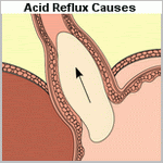
When the stomach contents reflux into the esophagus, its cellular lining is eroded. This damage mainly affects the superficial cells and the deep or basal germinative cells (stem cells) usually survive. In these cells a genetic switch causes them to generate new cells of specialized characteristics that differ from native esophageal cells. The new cells produce mucus and are more resistant to acid. They also differ in shape, being columnar instead of the spindle shaped esophageal cells. They also have some cells which are goblet shaped and are stuffed with mucus. This cellular pattern is similar to intestinal cells and is diagnostic of Barrett's esophagus.
About 10% of patients with acid reflux disease have Barrett's esophagus. It's usually diagnosed during endoscopy and has specific features.
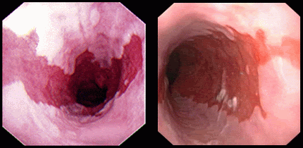
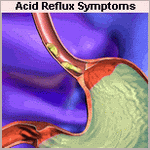
The pink projections characteristic of Barrett's esophagus vary in length, some are short (less than 3cm) and others long (more than 3cm) having higher risk of developing intestinal metaplasia . Proper diagnosis is confirmed by taking a specimen of this abnormal tissue and examining it under the microscope.
The process of replacement of esophageal cells with intestinal tissue (transformation of native cellular pattern with normal tissue of another organ) is called Metaplasia.
Metaplasia usually progresses into Dysplasia, a change in individual cellular features. Abnormalities involve cellular architecture, intracellular infrastructure and nuclei. Cells usually vary in size and shape and their nuclei reveal profound changes. Dysplasia may be low-grade or high-grade and this variety is pre-cancerous, usually complicated with a type of cancer called Adenocarcinoma of the esophagus. About 10% of patients with Metaplasia change into Dysplasia, and 1% of patients with Barrett's esophagus will have the risk of developing cancer.
As Barrett-type intestinal metaplastic cells are under-developed, they don't have normal sensory nerve supply and patients used to suffer from heartburn may not experience it any more. Accordingly, sufferers reporting spontaneous relief of symptoms after a long standing heartburn should be managed with a high index of suspicion.
In Barrett's esophagus It's mandatory to monitor cellular changes frequently. If no Dysplasia is associated endoscopy should be performed every year. When Dysplasia is detected, the opinion of an experienced pathologist is essential to confirm the diagnosis and differentiate between low-grade and high-grade types. In low-grade Dysplasia endoscopy should be repeated every six months to detect any progression. In high-grade Dysplasia, if the patient is at high risk for surgery, endoscopic ablative procedures should be considered. In fit patients surgical excision of the esophagus is the preferred approach, the operation is called esophagectomy.
Photo-dynamic therapy is an endoscopic ablative procedure which involves injecting a photo-sensitizing material followed by delivering red laser light to sensitized cells. Consequently, cells containing the drug are destroyed.
Endoscopic mucosal resection is safer than surgery and involves excision of dysplastic tissue, it also has a diagnostic role as it submits an adequate specimen for proper diagnosis and determining the depth of invasion.
Related posts:
Progress of acid reflux disease

