
TREATMENT OF ACID REFLUX COMPLICATIONS
Strictures
Mildly symptomatic esophageal strictures can be handled by careful attention to dietary intake, and use of medical therapy, primarily proton-pump inhibitors. Short, simple strictures can be dilated with weighted rubber or Teflon dilators (e.g., Hurst-Maloney). Tortuous or angulated strictures are more easily approached over a previously placed guidewire passed through an endoscope or under radiographic control (Savary dilators). Graded-steel olives (Eder-Puestow olive dilators), a dilator with graded increases of size (Celestin dilator), or a balloon with a fixed maximal diameter (Cooke balloon) can be passed over the previously placed wire. Alternatively, a balloon of fixed maximal diameter can be passed through the large channel of an endoscope during diagnostic endoscopy and dilated under direct vision (through-the-scope [TTS] dilation). Once the lumen is restored to a diameter of 13 to 15 mm, most patients swallow without difficulty. If the stricture is stable and requires dilation only every 4 to 6 months, no other therapy is necessary.
High-dose H2 -antagonists or, preferably, proton-pump inhibitors and dilation of the stricture can lead to healing of the mucosa and less need for repeated stricture dilation. Patients who do not tolerate dilation or require vigorous dilation every 3 to 4 weeks need a definitive antireflux operation, following which the stricture may regress. If strictures persist after antireflux surgery, esophageal replacement by colon, jejunum, or stomach is a surgical maneuver of last resort associated with a relatively high morbidity and mortality. Patients afflicted by strictures may have significant lung and cardiovascular disease that makes them unsuitable operative candidates.
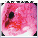
Ulcers
Esophageal ulcers also represent a major therapeutic problem. They usually require treatment with a proton-pump inhibitor.
Barrett's Esophagus
Barrett's (columnar) epithelium may be premalignant and can be removed only by esophageal resection. Adequate antireflux therapy with high-dose H2 -antagonists or with a proton-pump inhibitor causes regression of columnar epithelium in some patients.
Patients with Barrett's epithelium should be followed up with periodic endoscopic biopsies every 1 to 3 years to look for dysplasia and early changes of adenocarcinoma. The persistence of confirmed high-grade dysplasia is an indication for esophagectomy, because high-grade dysplasia may progress to carcinoma and because coexistent carcinoma may be undetected on biopsy. If low-grade dysplasia is present, the patient is treated medically with proton-pump inhibitors and undergoes biopsy every 6 to 12 months. Experimental endoscopic ablation therapies using photodynamic therapy, laser, or multipolar electrocoagulation are being tried to remove the columnar epithelium with the hope of subsequent growth of the normal squamous epithelium, primarily in patients with low-grade dysplasia or in patients with high-grade dysplasia who are not surgical candidates. Following these new ablation techniques, either long-term gastric acid suppression or laparoscopic Nissen fundoplication is needed to control the reflux and prevent recurrence of Barrett's epithelium.
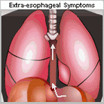
Pulmonary Complications
Treatment of the pulmonary complications of reflux in adults relies on improved night posture, gastric acid suppressants, and prokinetic agents. Caution is advised before recommending esophageal surgery in patients with reflux and predominant pulmonary problems, because the cause-and-effect relationship may be uncertain in individual patients.
Saturday, June 21, 2008
 TREATMENT OF ACID REFLUX COMPLICATIONS
Friday, May 16, 2008
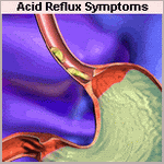
Acid Reflux Disease Complications
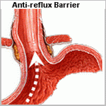
The complications of GE reflux result from the damage inflicted by gastric juice on the esophageal mucosa. Mucosal complications, include esophagitis and stricture. The prevalence and severity of complications is related to the degree of loss of the GE barrier, defects in esophageal clearance, and the content of refluxed gastric juice.
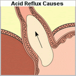
The potential injurious components that reflux into the esophagus include gastric secretions, such as acid and pepsin, biliary and pancreatic secretions that regurgitate from the duodenum into the stomach, and toxic compounds generated in the mouth, esophagus, and stomach by the action of bacteria on dietary substances.
Studies have shown that acid alone does minimal damage to the esophageal mucosa, but the combination of acid and pepsin is highly deleterious. Hydrogen ion injury to the esophageal squamous mucosa occurs only at a pH below 2. In acid refluxate, the enzyme pepsin appears to be the major injurious agent. Similarly, the reflux of duodenal juice alone does little damage to the mucosa, whereas the combination of duodenal juice and gastric acid is particularly noxious. Reflux of bile and pancreatic enzymes into the stomach can either protect against or augment esophageal mucosal injury. For instance, the reflux of duodenal contents into the stomach may prevent the development of peptic esophagitis in a patient whose gastric acid secretion maintains an acid environment, because the bile salts would attenuate the injurious effect of pepsin and the acid would inactivate the trypsin. Such a patient would have bile-containing acid gastric juice that, when refluxed, would irritate
the esophageal mucosa but cause less esophagitis than if it were acid gastric juice containing pepsin. In contrast, the reflux of duodenal contents into the stomach of a patient with limited gastric acid secretion can result in esophagitis, because the alkaline intragastric environment would support optimal trypsin activity, and the soluble bile salts with a high pKa would potentiate the enzyme's effect. Hence, duodenal-gastric reflux and the acid secretory capacity of the stomach interrelate by altering the pH and enzymatic activity of the refluxed gastric juice to modulate the injurious effects of enzymes on the esophageal mucosa.
This disparity in injury caused by acid and bile alone, as opposed to the gross esophagitis caused by pepsin and trypsin, provides an explanation for the poor correlation between the symptom of heartburn and endoscopic esophagitis. The reflux of acid gastric juice contaminated with duodenal contents could break the esophageal mucosal barrier, irritate nerve endings in the papillae close to the luminal surface, and cause severe heartburn. Despite the presence of intense heartburn, the bile salts present would inhibit pepsin, the acid pH would inactivate trypsin, and the patient would have little or no gross evidence of esophagitis. In contrast, the patient who refluxed alkaline gastric juice may have minimal heartburn because of the absence of hydrogen ions in the refluxate but have endoscopic esophagitis because of the bile salt potentiation of trypsin activity on the esophageal mucosa. This is supported by recent clinical studies which indicate that the presence of alkaline reflux is associated with the development of mucosal injury.
Although numerous studies have suggested the reflux of duodenal contents into the esophagus in patients with GERD, few have measured this directly. The components of duodenal juice thought to be most damaging are the bile acids and, as such, they have been the most commonly studied. Most studies shown that, patients with GERD have greater and more concentrated bile acid exposure to the esophageal mucosa than do normal subjects. This increased exposure occurs most commonly during the supine period while asleep and during the upright period following meals. Most studies have identified the glycine conjugates of cholic, deoxycholic, and chenodeoxycholic acids as the predominant bile acids aspirated from the esophagus of patients with GERD, although appreciable amounts of taurine conjugates of these bile acids were also found. Other bile salts were identified but in small concentrations. This is as one would expect because glycine conjugates are three times more prevalent than taurine conjugates in normal human bile.
The potentially injurious action of toxic compounds either ingested or newly formed on the mucosa of the GE junction and distal esophagus has long been postulated. Investigators have recently shown that dietary nitrate consumed in the form of green vegetables and food contaminated by nitrate-containing fertilizers results in the generation of nitric oxide at the GE junction in concentrations high enough to be potentially mutagenic. Previous studies have shown that nitrate ingested in food is reabsorbed in the small bowel with approximately 25% resecreted into the mouth via the salivary glands. Oral bacteria chemically transform the relatively innocuous nitrate to the more toxic nitrite, which is swallowed and subsequently converted to nitric oxide and other toxic nitroso-compounds by acid and ascorbic acid in the stomach. Whether this mechanism in fact contributes to injury and or neoplastic transformation in the upper stomach, GE junction, and distal esophagus is currently unknown.

