
The development of fibre-optic endoscopy revolutionized the ability to investigate the gastrointestinal tract. Flexible endoscopes often have a diameter of less than 1 cm, with a control head and a flexible shaft with a manoeuvrable tip. The head is connected to a light source and can transmit images to a video image screen.
All endoscopes have multiple small lumens allowing transmission of air and water and for suction. The suction channel can also be used for the passage of interventional devices, for example biopsy forceps. The ability to transmit air (insufflation) allows the endoscopist to inflate the lumen to obtain optimal views. The water channel provides a means of washing mucosal surfaces, and suction maybe used to remove pools of fluid within the gastrointestinal tract, thus ensuring that all mucosal surfaces are inspected.
Diagnostic indications include the investigation of gastro-esophageal reflux (diagnostic investigation or surveillance endoscopy of Barrett's esophagus).
Therapeutic procedures that can be undertaken include mucosal biopsy, stent insertion for strictures, and dilatation of strictures.
Patient preparation
Informed consent is required, and patients are fasted for 4-6 hours prior to the procedure. Although most patients do not require sedation, the choice is offered and discussed. Sedation involves the use of a short-acting benzodiazepine which provides a sedative and amnesic effect. Monitoringis required (pulse oximetry) with sedation due to the risk of respiratory depression. Antibiotic prophylaxis is administered to patients with heart valve disease to prevent bacterial endocarditis.
Procedure
A mouthguard is used. The endoscope is introduced into the pharynx, then the oesophagus. Patients may retch during this procedure. The endoscope is progressively introduced to inspect the oesophagus, stomach and proximal small bowel (duodenum).
Complications
The overall complication rate is approximately 1 per 1000 with a mortality rate of approximately 1 per 25000. Complications include bleeding, perforation and respiratory arrest (a complication of sedation).
Post procedure care
Patients are monitored in a recovery area until safe for discharge.
Saturday, May 3, 2008
 Endoscopic investigation of gastro-esophageal reflux
Friday, March 7, 2008
Endoscopic evaluation
Endoscopic visualization of the esophagus equates to the physical examination of the foregut and is a critical part of the preoperative evaluation of patients with GERD. Its main aim is to detect complications of GE reflux, the presence of which may influence therapeutic decisions.
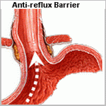
In every patient, the locations of the diaphragmatic crura, the GE junction, and the squamocolumnar junction are determined. These anatomic landmarks are commonly at three different sites in patients with GERD. The crura are usually evident and can be confirmed by having the patient sniff during the examination. The anatomic GE junction is identified as the point where the gastric rugal folds meet the tubular esophagus and is often below the squamocolumnar junction, even in patients without otherwise obvious Barrett's esophagus.
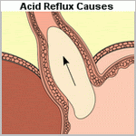
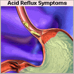
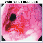
Endoscopic esophagitis is defined by the presence of mucosal erosions . When present, the grade and length of esophageal mucosal injury are recorded. The presence and length of columnar epithelium extending above the anatomic GE junction is also noted. It is suspected at endoscopy when there is difficulty in visualizing the squamocolumnar junction at its normal location and by the appearance of a velvety red luxuriant mucosa. The presence of Barrett's esophagus is confirmed by biopsy evidence of specialized intestinal metaplasia and is considered histologic evidence of GERD. Endoscopic visualization of columnar lining without histologic confirmation of specialized intestinal metaplasia is not considered Barrett's esophagus and likely has no premalignant potential. Multiple biopsies should be taken in a cephalad direction to determine the level at which the junction of Barrett's epithelium and normal squamous mucosa occurs. Barrett's esophagus is susceptible to ulceration, bleeding, stricture formation, and malignant degeneration. Dysplasia is the earliest sign of malignant change. Because dysplastic changes typically occur in a random distribution within the distal esophagus, a minimum of four biopsies (each quadrant) every 2 cm should be obtained from the metaplastic epithelium. Particular attention must be paid to the squamocolumnar junction in these patients, where a mass, ulcer, nodularity, or inflammatory tissue is always considered suspicious for malignancy and requires thorough biopsy. The GE junction is defined endoscopically where the tubular esophagus meets gastric rugal folds, and the squamocolumnar junction is where there is an obvious change from the velvety and darker columnar epithelium to the lighter squamous epithelium.
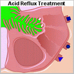
After completion of the esophageal examination, the first and second portions of the duodenum and the stomach are systematically inspected. This is commonly done on withdrawal of the endoscope. When the antrum is visualized, the incisura angularis appears as a constant ridge on the lesser curve. Turning the lens of the scope 180 degrees allows inspection of the fundus and cardia. Attention is paid to the frenulum (angle of His) of the esophagogastric junction and to the closeness with which the cardia grips the scope. The appearance of this valve have been graded on a scale from I to IV according to the degree of unfolding or deterioration of the normal valve architecture. This grading system has been correlated with the presence of increased esophageal acid exposure, occurring predominantly in patients with a grade III or IV valve.
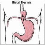
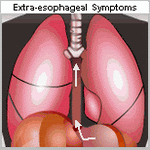
A hiatal hernia is endoscopically confirmed by finding a pouch lined with gastric rugal folds lying 2 cm or more above the margins of the diaphragmatic crura. A prominent sliding hernia is frequently associated with increased esophageal exposure to gastric juice. When a paraesophageal hernia exists, particular attention is given to exclude a gastric ulcer or gastritis within the pouch. The intragastric retroflex or J maneuver is important in evaluating the full circumference of the mucosal lining of the herniated stomach. As the endoscope is removed, the esophagus is again examined and biopsies taken. The location of the cricopharyngeus is identified and the larynx and vocal cords are visualized. Acid reflux may result in inflammation of the larynx. Vocal cord movement is recorded both as a reference for subsequent surgery and an assessment of the patient's ability to protect the airway.

