
 Radiographic evaluation
Radiographic assessment of the anatomy and function of the esophagus and stomach is one of the most important parts of the preoperative evaluation. Critical issues are assessed, including the presence of esophageal shortening, the size and reducibility of a hiatal hernia, and the propulsive function of the esophagus for both liquids and solids.
The definition of radiographic GE reflux varies depending on whether reflux is spontaneous or induced by various maneuvers. In only about 40% of patients with classic symptoms of GERD is spontaneous reflux observed by the radiologist (i.e., reflux of barium from the stomach into the esophagus with the patient in the upright position). In most patients who show spontaneous reflux on radiography, the diagnosis of increased esophageal acid exposure is confirmed by 24-hour esophageal pH monitoring. Therefore, the radiographic demonstration of spontaneous regurgitation of barium into the esophagus in the upright position is a reliable indicator that reflux is present. Failure to see this does not indicate the absence of disease.
A carefully performed video esophagram can provide an enormous amount of information on the structure and function of the esophagus and stomach. The modern barium swallow emphasizes motion-recording (video), utilizes a tightly controlled examination protocol, and requires an understanding of esophageal physiology.
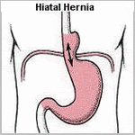
Videotaping the study greatly aids the evaluation, providing the surgeon with a real-time assessment of swallowing function, bolus transport, and the size and reducibility of hiatal hernias. Given routine review before antireflux surgery, its value becomes increasingly clear. The study provides structural information including the presence of obstructing lesions and anatomic abnormalities of the foregut. A hiatal hernia is present in more than 80% of patients with GE reflux and is best demonstrated with the patient in the prone position, which causes increased abdominal pressure and promotes distention of the hernia above the diaphragm. The presence of a hiatal hernia is an important component of the underlying pathophysiology of GE reflux. Other relevant findings include a large (greater than 5 cm) or irreducible hernia, suggesting the presence of a shortened esophagus; a tight crural collar that inhibits barium transit into the stomach, suggesting a possible cause of dysphagia; and the presence of a paraesophageal hernia.
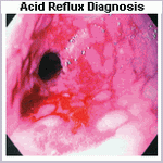
Lower esophageal narrowing resulting from a ring, stricture, or obstructing lesion is optimally viewed with full distention of the esophagogastric region. A full-column technique with distention of the esophageal wall can be used to discern extrinsic compression of the esophagus. Mucosal relief or double-contrast films should be obtained to enhance the detection of small esophageal neoplasms, mild esophagitis, and esophageal varices. The pharynx and upper esophageal sphincter are evaluated in the upright position, and an assessment of the relative timing and coordination of pharyngeal transit is possible.
The assessment of peristalsis on video esophagram often adds to, or complements, the information obtained by esophageal motility studies. This is in part because the video barium study can be done both upright and supine and with liquid and solid bolus material, which is not true of a stationary motility examination. This is particularly true with subtle motility abnormalities. During normal swallowing, a stripping wave (primary peristalsis) is generated that completely clears the bolus. Residual material can stimulate a secondary peristaltic wave, but usually a second pharyngeal swallow is required. Motility disorders with disorganized or simultaneous esophageal contractions have tertiary waves and provide a segmented appearance to the barium column, often referred to as beading or corkscrewing. In dysphagic patients, a barium-impregnated marshmallow, bread, or hamburger is a useful adjunct, which can discern a functional esophageal transport disturbance not evident on the liquid barium study. Reflux is not easily seen on video esophagram, and motility disorders that cause retrograde barium transport may be mistaken for reflux.
Assessment of the stomach and duodenum during the barium study is a necessity for proper preoperative evaluation of the patient with GERD. Evidence of gastric or duodenal ulcer, neoplasm, or poor gastroduodenal transit has obvious importance in the proper preoperative evaluation.
skip to main |
skip to sidebar


Acid Reflux Relief
Acid Reflux Relief: TOC
Loading...
Acid Reflux Relief: Subscribe
Acid Reflux Relief: Archive
-
▼
2008
(36)
-
▼
March
(15)
- GERD Asthma Treatment
- GERD Asthma
- What is (GERD)?
- Great Videos about Acid Reflux and its Relief
- How common is Acid Reflux?
- Medications for Acid Reflux Relief
- Assessment of esophageal body and gastric function
- Radiographic evaluation
- Assessment of esophageal length
- Twenty-four hour ambulatory pH monitoring
- Endoscopic evaluation
- Surgery for Acid Reflux Relief
- Acid Reflux Symptoms
- Acid Reflux (GERD) Videos
- Acid Reflux (Heartburn) Videos
-
▼
March
(15)
Copyright © 2007 Acid Reflux Relief